| 
  • If you are citizen of an European Union member nation, you may not use this service unless you are at least 16 years old.

  • You already know Dokkio is an AI-powered assistant to organize & manage your digital files & messages. Very soon, Dokkio will support Outlook as well as One Drive. Check it out today!

View
 

Hyperglycemic Crises: Diabetic Ketoacidosis (DKA), And Hyperglycemic Hyperosmolar State (HHS)

Page history last edited by Dhemy Padilla 14 years, 6 months ago


The Web diabetesmanager


HYPERGLYCEMIC CRISES IN PATIENTS WITH DIABETES: DIABETIC KETOACIDOSIS (DKA), AND HYPERGLYCEMIC HYPEROSMOLAR STATE (HHS)


Last Author Revision:  2009 


Introduction

Diabetic ketoacidosis (DKA) and hyperosmolar hyperglycemic state (HHS) represent two extremes in the spectrum of marked decompensated diabetes. DKA and HHS are still important causes of morbidity and mortality among patients with diabetes even with major agreements about their diagnostic criteria and treatment protocols [1][2]. The annual incidence of DKA from population-based studies is estimated to range from 4 to 8 episodes per 1,000 patient admissions with diabetes [3]. The incidence of DKA continues to increase with DKA accounting for about 115,000 hospitalizations in the United States in 2003 (figure 1 a)[4]. The rate of hospital admissions for HHS is lower than DKA and is less than 1% of all diabetic-related admissions [5][6][7][8]. Decompensated diabetes imposes a heavy burden in terms of economics and patient outcomes. DKA is an economically burdensome with the average cost of $13,000 per patient per hospitalization [9].Therefore, the annual expenditure for the care of patients with DKA may exceed $1 billion. The mortality rate for DKA has been falling over the years. Age adjusted mortality rates in the U.S. have dropped by 22% between 1980 and 2001 (from 32 to 20 per 100,000 diabetic population respectively)(4)(figure 1b). Contrary to DKA mortality, the mortality rate of HHS has remained high, ~ 15%, compared to less than 5% in patients with DKA [10][11][12][13][14]. Severe dehydration, older age, and the presence of comorbid conditions in patients with HHS, account for the higher mortality in these patients [15].


Figure 1a. Incidence of DKA 1980-2003  [16]


Figure 1b. Mortality rate of DKA 1980-2001 [17] 


Definitions

DKA consists of the biochemical triad of hyperglycemia, ketonemia and metabolic high anion gap acidosis [18] (Figure 2). The terms “hyperglycemic hyperosmolar nonketotic state” and “hyperglycemic hyperosmolar nonketotic coma” have been replaced with the term “hyperglycemic hyperosmolar state” (HHS) [19] to show the facts that 1)the hyperglycemic hyperosmolar state may consist of moderate to variable degrees of clinical ketosis detected by nitroprusside method and 2) alterations in consciousness may often be present without coma .


Figure 2. The triad of DKA (hyperglycemia, acidemia, and ketonemia) and other conditions with which the individual components are associated. From Kitabchi and Wall [20]


Both DKA and HHS are characterized by absolute or relative insulinopenia. Clinically, they differ only by the severity of dehydration, ketosis and metabolic acidosis [21][22].


DKA most often occurs in patients with type 1 diabetes mellitus (T1DM). It also occurs in type 2 diabetes under conditions of extreme stress such as serious infection, trauma, cardiovascular or other emergencies, and, less often, as a presenting manifestation of type 2 diabetes, a disorder called ketosis-prone type 2 diabetes [23][24]. Similarly, whereas HHS occurs most commonly in T2DM, it can be seen in T1DM in conjunction with DKA [25][26].


Pathogenesis

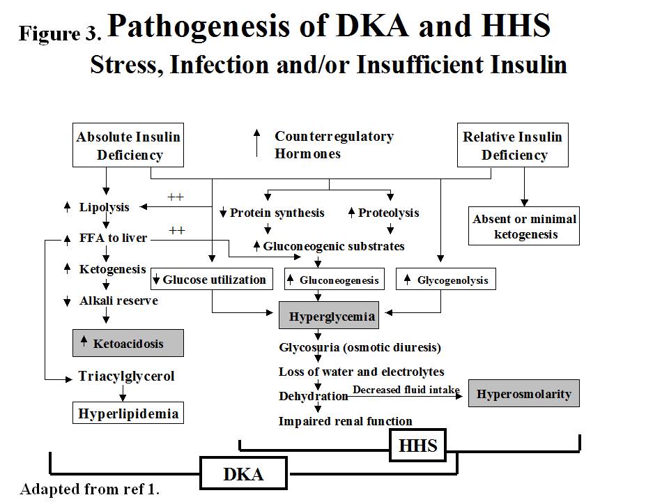
The underlying defects in DKA and HHS are 1) reduced net effective action of circulating insulin as a result of decreased insulin secretion (DKA) or ineffective action of insulin in HHS [27][28][29] 2)elevated levels of counterregulatory hormones: glucagon [30][31], catecholamines [32][33], cortisol [34][35], and growth hormone [36][37], resulting in increased hepatic glucose production and impaired glucose utilization in peripheral tissues 3)dehydration and electrolytes abnormalities mainly due to osmotic diuresis caused by glycosuria [38][39](figure 3).


Figure 3. Pathogenesis of DKA and HHS


Diabetic Ketoacidosis

In DKA, there is severe alteration of carbohydrate, protein, and lipid metabolism (1). In general, the body is shifted into a major catabolic state with breakdown of glycogen stores, hydrolysis of triglycerides from adipose tissues and mobilization of aminoacids from muscle [40].The released triglycerides and amino acids from the peripheral tissues will be the substrates for the production of glucose and ketone bodies by the liver [41] .Hyperglycemia and ketone bodies production play central roles in developing this metabolic decompensation [42].


Hyperglycemia

The hyperglycemia in DKA is the result of three events: (a) increased gluconeogenesis; (b) increased glycogenolysis, and (c) decreased glucose utilization by liver, muscle and fat. Decreased insulin and elevated cortisol levels also result in decreased protein synthesis and increased proteolysis with increased production of amino acids (alanine and glutamine), which serve as substrates for gluconeogenesis [43][44]. Furthermore, muscle glycogen is catabolized to lactic acid via glycogenolysis. The lactic acid is transported to the liver in the Cori cycle where it serves as carbon skeleton for gluconeogenesis [45]. Increased levels of glucagon, cathecholamines and cortisol with concurrent insulinopenia stimulate gluconeogenic enzymes especially phosphoenol pyruvate carboxykinase (PEPCK) [46][47]. Decreased glucose utilization is further exaggerated by increased levels of circulating catecholamines and FFA [48].


Ketogenesis

Excess catecholamines coupled with effective insulinopenia promote triglyceride breakdown (lipolysis) to free fatty acids (FFA) and glycerol, the latter provides carbon skeleton for gluconeogenesis while the former provides the substrate for the formation of ketone bodies [49][50]. The key regulatory site for fatty acid oxidation is known to be carnitine palmitoyltransferase 1(CPT1)which is inhibited by malonyl CoA in normal nonfasted state but the increase ratio of glucagons and other counter regulatory hormones to insulin disinhibit the fatty acid oxidation and the incoming fatty acids from fat tissue can be converted to ketone bodies [51][52]. Increased production of ketone bodies (acetoacetate, and ß- hydroxybutyrate) leads to ketonemia [53]. There is also decreased clearance of ketone bodies in DKA, which contributes to ketonemia [54]. These ketoacids are buffered by extracellular and cellular buffers resulting in their loss and subsequent anion gap metabolic acidosis [55]. Studies in diabetic and pancreatectomized patients have demonstrated the cardinal role of hyperglucagonemia and insulinopenia in the genesis of DKA [56](Figure 4).. In the absence of stressful situations such as dehydration, vomiting or intercurrent illness, ketosis is usually mild [57][58].


Figure 4. Proposed Biochemical Alterations in Diabetic Ketoacidosis Leading to Increased Gluconeogenesis, Lipolysis, Ketogenesis and Decreased Glycolysis.  [59]


Elevated levels of pro-inflammatory cytokines and lipid peroxidation markers, as well as procoagulant factors such as plasminogen activator inhibitor-1 (PAI-1) and C-reactive protein (CRP) have been demonstrated in DKA. The levels of these factors return to normal with insulin therapy and correction of hyperglycemia [60]. This inflammatory and procoagulant state may explain the well-known association between hyperglycemic crisis and thrombotic state.


Hyperglycemic Hyperosmolar State

The pathogenesis of DKA and HHS are similar, however, in HHS: 1) there is enough insulin to prevent lipolysis and ketogenesis but not adequate to cause glucose utilization (as it takes 1/10 as much insulin to suppress lipolysis as it does to stimulate glucose utilization)(47,48) 2) possible smaller increases in counterregulatory hormones [61][62]


Precipitating factors

The two most common precipitating factors in the development of DKA or HHS are inadequate or inappropriate insulin therapy or infection [63][64]. Other factors include myocardial infarction, cerebrovascular accidents,pulmonary embolism, pancreatitis ,alcohol abuse and drugs [65] (Table 1) . In addition to the mentioned precipitating factors, numerous underlying medical illness and medications that provoke the release of counter regulatory hormones and/or compromise the access to water are likely to result in severe dehydration and HHS [66]. Drugs such as corticosteroids, thiazides, sympathomimetic agents (e.g.,dobutamine and terbutaline ) [67] and second generation antipsychotic agents [68] may precipitate the genesis of DKA or HHS. In young patients with type 1 diabetes, insulin omission due to fear of hypoglycaemia or weight gain, the stress of chronic disease, and eating disorders, may contribute in 20 % of recurrent DKA [69]. Cocaine use also is associated with recurrent DKA [70][71]. Mechanical problems with continuous subcutaneous insulin infusion (CSII) devices had also precipitated DKA [72], but with improvement in technology and better education of patients ,the incidence of DKA seems to have been reduced in pump users [73]. Further studies are required to document reduction of DKA incidence with the use of CSII devices. There are also case reports of patients with DKA as the primary manifestation of acromegaly [74][75][76][77].


Table 1. Diagnostic Criteria and Typical Total Body Deficits of Water and Electrolytes in Diabetic Ketoacidosis (DKA) and Hyperglycemic Hyperosmolar Syndrome (HHS)


Increasing numbers of DKA cases have been reported in patients with DMT2. Available evidence shows that almost 50 % of newly diagnosed adult African American and Hispanic patients with DKA have type 2 diabetes [78].These patients who have ketosis– prone type 2 diabetes develop sudden-onset impairment in insulin secretion and action, resulting in profound insulinopenia [79]. Clinical and metabolic features of these patients include a high rate of obesity, a strong family history of diabetes, a measurable pancreatic insulin reserve and low prevalence of autoimmune markers of ß-cell destruction  [80][81] .Aggressive management with insulin improves ß cell function , leading to discontinuance of insulin therapy within a few months of follow-up and 40 % of these patients remain non insulin dependent for 10 years after the initial episode of DKA  [82][83].The etiology of acute transient failure of ß-cells leading to DKA in these patients is not known but the suggested mechanisms include glucotoxicity, lipotoxicity, and genetic predisposition [84][85].


A genetic disease, glucose-6-phosphate dehydrogenase deficiency, has been linked with ketosis-prone diabetes  [86].

Table 2 sumarizes the common precipitating factors in DKA. 


Table 2. Precipitating factors for DKA


Clinical Features


Symptoms and signs

DKA evolves rapidly within a few hours of the precipitating event(s). On the other hand, development of HHS is insidious and may occur over days or weeks [87].


The common clinical pictures in DKA and HHS due to hyperglycemia include polyuria, polyphagia, polydipsia, weight loss, weakness and physical signs of dehydration such as dry buccal mucosa, sunken eye balls, poor skin turgor, tachycardia, hypotension and shock in severe cases. Kussmaul respiration, acetone breath, nausea, vomiting and abdominal pain may also occur primarily in DKA . Abdominal pain, which correlates with the severity of acidosis [88], may be severe enough to be confused with acute abdomen in 50-75% of cases [89]. Therefore, in the presence of acidosis, DKA as an etiology of abdominal pain should be considered [90]. Patients often are normothermic or mild hypothermia, even when harbouring an infection [91].Therefore careful search for a source of infection should be performed even in the absence of fever [92]. Mental status in DKA may vary from full alertness to profound lethargy or coma but less frequent than HHS [93][94]. The relationship of depressed consciousness to higher serum osmolality in DKA and its causes has been controversial [95][96], some studies have suggested that pH is its cause [97] while others have concluded that osmolality [98] is responsible for the comatose state. More recently it has been proposed that consciousness level in children with DKA is related to the severity of acidosis (pH) but not to blood glucose [99].  This however was reported in children under the age of 16. In our earlier studies of patients with DKA using low dose versus high dose insulin therapy, we evaluated the initial biochemical values of 48 patients with stupor/coma versus non comatose patients [100]. Table 3 shows that only glucose, bicarbonate, BUN and osmolality, but not pH , were significantly different between non comatose and comatose patients. Furthermore in 3 separate studies in which 123 cases of DKA were evaluated, the most significant finding was related to osmolality as demonstrated in figure 5 [101]. This finding was also repeated in other study with different patients which showed osmolality was related significantly to glucose levels but not to pH [102]. Furthermore ,according to a recent study, ICU-admitted patients with DKA are less ill, and have lower disease severity scores, mortality, and shorter length of ICU and hospital stay, than non-DKA patients. Disease severity scores are not, but precipitating cause is, predictive of prolonged hospital stays in patients with DKA [103]


Figure 5. Relationship between serum osmolality and level of consciousness. (Adapted from ref [104])


Table 3. Admission clinical and biochemical profile and response to therapy of comatose versus noncomatose patients with DKA.  [105]


In patients with HHS, final symptoms include clouding of sensorium which progresses to mental obtundation or coma [106]. Occasionally, patients with HHS may present with focal neurological deficit and seizure disorders [107][108]. Most of the patients with HHS and an effective osmolarity of > 320 mOsm/kg are severely obtundated or comatose, but altered mental status rarely exists in patients with osmolarity of < 320 mOsm/kg [109]  (Figure 5).Therefore, severe alteration in the level of consciousness in patients with osmolarity of < 320 mOsm/kg requires evaluation for other causes including CVA and other catastrophic events like myocardial and bowel infarctions   [110][111].


Laboratory Abnormalities

The initial laboratory evaluation of patients with suspected DKA or HHS should include determination of plasma glucose, blood urea nitrogen, creatinine , serum ketones, electrolytes (with calculated anion gap), osmolality, urinalysis, urine ketones by dipstick, arterial blood gases and complete blood count with differential. An electrocardiogram, blood, urine or sputum cultures and chest X-ray should also be performed, if indicated. HbA1c may be useful in differentiating chronic hyperglycemia of uncontrolled diabetes from acute metabolic decompensation in a previously well controlled diabetic patient [112]. Table 1 summarizes the biochemical criteria for DKA and HHS and electrolyte deficits in these two conditions. It also provides a simple method for calculating anion gap and serum osmolality.[113]


DKA is classified as mild, moderate, or severe based on the severity of metabolic acidosis and the presence of altered mental status [114][115]. Over 30% of patients have features of both DKA and HHS [116]. Patients with HHS typically have pH > 7.30, bicarbonate level > 20 mEq/L, and negative ketone bodies in plasma and urine. However, some of them may have ketonemia. Some studies on serum osmolarity and mental alteration have established a positive linear relationship between osmolarity and mental obtundation [117][118].  Therefore , the occurrence of coma in the absence of definitive elevation of serum osmolality requires immediate consideration of other causes of mental status change [119][120].


The admission biochemical data in patients with DKA or HHS are shown in Table 4.


Table 4. Admission biochemical data in patients with HHS and DKA.


The major cause of water deficit in DKA and HHS is glucose-mediated osmotic diuresis which leads to loss of water in excess of electrolytes [121]. Despite the excessive water loss, the admission serum sodium tends to be low because serum glucose in the presence of insulinopenia of DKA and HHS cannot penetrate to cells. Therefor it becomes osmotically effective and pulls water from intracellular space to the extra cellular space and dilutes the sodium concentration [122]. True sodium concentration (millimolar )can be obtained by multiplying excess glucose above 100 mg/dl by 1.6 /100 [123].  If the corrected sodium level is extremely low , hypertriglyceridemia (secondary to uncontrolled diabetes ) should be suspected. In this condition the plasma becomes milky and lipema retinalis may be visible in physical examination [124]. Serum potassium may be elevated on arrival due to insulin deficiency, dehydration and a shift of potassium from intracellular to extra cellular compartments in response to acidosis [125]. On occasion, the initial potassium level may be normal or low, which is a danger sign Initiation of therapy, which leads to the transfer of potassium into cells, may cause fatal hypokalemia if potassium is not added early [126].  Phosphate depletion in DKA is universal but on admission, like the potassium, it may be low ,normal or high [127].


Leukocytosis is a common finding in patients with DKA or HHS [128][129], but leukocytosis greater than 25,000 /μL suggests ongoing infection requiring further work up [130]. The exact etiology of this non specific leukocytosis is not known. A recent study also showed  non specific leukocytosis in subjects with hyoglycaemia induced by insulin injection and suggested that this phenomenon may be due to the increased levels of cathecholamine, cortisol, and proinflammatory cytokines such as TNF-α during acute stress [131]. Hypertriglyceridemia may be present in HHS  [132] and is almost always seen in DKA [133].  Hyperamylasemia, which correlates with pH and serum osmolality and elevated level of lipase may occur in 16 - 25% of patients with DKA [134]. The origin of amylase in DKA is usually non-pancreatic tissue such as the parotid gland [135].


Pitfalls of Laboratory Tests

False positive values for lipase may also be seen if plasma glycerol levels are very high due to rapid breakdown of adipose tissue triglycerides (glycerol is the product measured in most assays for plasma lipase). Therefore, pancreatic enzymes may not be reliable tools for the diagnosis of pancreatitis in the setting of DKA [136][137]. Other pitfalls include artificial elevation of serum creatinine, either as a result of dehydration or interference from ketone bodies if a colorimetric method is used [138]. Most of the laboratory tests for ketone bodies use the nitroprusside method, which detects acetoacetate, but not β hydroxybutyrate (BOHB ) [139]. Since BOHB is converted to acetoacetate during treatment [140], the ketone test may show high values suggesting erroneously that ketonemia is deteriorating, therefore the follow up measurement of ketones during the treatment by nitroprusside method is not recommended [141].Newer glucose meters have the capability to measure BOHB, which overcomes this problem  [142][143],( Figure 6 ). Furthermore drugs that have sulfhydryl groups can interact with the reagent in the nitroprusside reaction, giving a false positive result [144]. Particularly important in this regard is captopril, an angiotensin converting enzyme inhibitor prescribed for the treatment of hypertension and diabetic nephropathy. Therefore for the diagnosis of DKA, clinical judgement and consideration of other biochemical data are required to interpret the value of positive nitroprusside reactions in patients on captopril [145].


Figure 6. Comparative data in 37 patients with diabetic ketoacidosis with regard to plasma acetoacetic acid (AA) and -hydroxybutyric acid ( OH) (top left); ratio of AA to OH (bottom left); and ketone bodies (nitroprusside reaction) in the urine (top right) and plasma (bottom right) before and during low-dose intravenous infusion of insulin for 48 hours. Reproduced with permission from Reference [146].


DIFFERENTIAL DIAGNOSIS

Patients may present with metabolic conditions resembling DKA or HHS. For example, in alcoholic ketoacidosis (AKA), total ketone bodies are much greater than in DKA with a higher BOHB to acetoacetate ratio of 7:1 versus a ratio of 3:1 in DKA [147][148]. The AKA patients seldom present with hyperglycemia [149]. It is also possible that patients with a low intake of food will present with mild ketoacidosis (starvation ketosis) but these patients rarely present with serum bicarbonate concentration less than 18, and do not exhibit hyperglycemia [150]. Additionally, DKA has to be distinguished from other causes of high anion gap acidosis including lactic acidosis, advanced chronic renal failure as well as ingestion of drugs such as salicylate, methanol and ethylene glycol. Isopropyl alcohol, which is commonly available as rubbing alcohol can cause considerable ketosis and high osmolar gap without metabolic acidosis, but it has a tendency to cause hypoglycemia rather than hyperglycemia [151][152]. Finally, patients with diabetes insipidus presenting with severe polyuria and dehydration, who are treated with dextrose water, can have hyperglycemia ; a clinical picture that can be confused with HHS [153], (Table 5).


Table 5. Laboratory evaluation of metabolic causes of acidosis and coma


TREATMENT

The goals of therapy in patients with DKA and HHS include 1) Improvement of circulatory volume and tissue perfusion, 2) Gradual reduction of serum glucose and plasma osmolarity, 3) correction of electrolyte imbalance, and in DKA steady resolution of ketosis, and 4) Identification and prompt treatment of co-morbid precipitating causes [154][155]. It must be emphasized that successful treatment of DKA and HHS requires frequent monitoring of patients regarding the above goals by clinical and laboratory parameters. Protocols for the management of patients with DKA and HHS are illustrated in figures 7 . A flow sheet such as the one shown in figure 8 should be provided for monitoring the patients [156].


Figure 7. Protocol for the management of adult patients with DKA and HHS.


*DKA diagnostic criteria: serum glucose >250 mg/dl, arterial pH <7.3, serum bicarbonate <18 mEq/l, and moderate ketonuria or ketonemia. Normal laboratory values vary; check local lab normal ranges for all electrolytes. After history and physical exam, obtain capillary glucose and serum or urine ketones (nitroprusside method). Begin 1 liter of 0.9% NaCl over 1 h and draw arterial blood gases, complete blood count with differential, urinalysis, serum glucose, BUN, electrolytes, chemistry profile, and creatinine levels STAT. Obtain electrocardiogram, chest X-ray, and specimens for bacterial cultures, as needed. *Serum Na+ should be corrected for hyperglycemia (for each 100 mg/dl glucose >100 mg/dl, add 1.6 mEq to sodium value for corrected serum sodium value). Adapted from ref. [157].

 **HHS diagnostic criteria: serum glucose > 600 mg/dl, arterial pH >7.3, serum bicarbonate > 15 Meq/l, and minimal ketonuria and ketonemia. Normal laboratory values vary; check local lab normal ranges for all electrolytes. † After history and physical exam, obtain capillary glucose and serum or urine ketones (nitroprusside method). Begin 1 liter of 0.9% NaCl over 1h and draw arterial blood gases, complete blood count with differential urinalysis, serum glucose, BUN, elecrolytes, chemistry profile and creatinine levels STAT. Obtain elecrocardiogram, chest X-ray, and specimens for bacterial cultures, as needed. Adapted from ref. [158] * Serum Na+ should be corrected for hyperglycemia (for each 100 mg/dl gludose > 100 mg/dl, add 1.6 mEq to sodium value for corrected serum sodium value).


Fluid Therapy

DKA and HHS are volume-depleted states with water deficit of approximately 6 L in DKA and 9 L in HHS [159][160]. Therefore, the initial fluid therapy is directed toward expansion of interstitial and intravascular volume and securing adequate urine flow . The initial fluid of choice is isotonic saline, which we recommend to be infused at the rate of 15–20 ml /kg body weight per hour or 1–1.5 L during the first hour. The choice of fluid for continued repletion depends on the hydration status, serum electrolyte levels, and urinary output. In patients who are hypernatremic or eunatremic, 0.45% NaCl infused at 4–14 ml/kg/hour is appropriate and in patients with hyponatremia 0.9% NaCl at a similar rate is preferred. The goal is to replace half of the estimated water deficit over a period of 12- 24 hours [161]. In patients with hypotension, aggressive fluid therapy with isotonic saline should continue until blood pressure is stabilized. The administration of insulin without fluid replacement in such patients may further aggravate hypotension [162].  Furthermore, the use of hydrating fluid in the first hour of therapy before insulin has multiple advantages including a) providing a chance to obtain serum potassium value before insulin administration, b) preventing possible deterioration of hypotensive patients with the use of insulin without adequate hydration, and c) increasing insulin effectiveness by decreasing the serum osmolality [163][164]. Hydration alone may also reduce the level of counterregulatory hormones and hyperglycemia [165][166]. Hydration reduces serum blood glucose, BUN, and potassium levels without significant changes in pH or HCO3 [167].The mechanism for lowering glucose is believed to be due to osmotic diuresis and modulation of conterregulatory hormone release [168][169]. Patients with DKA and HHS require calories for proper metabolism of ketone bodies. Therefore in DKA, as soon as blood glucose falls below 200 mg/dl, the sodium chloride solution should be replaced with 5% glucose containing saline solution with a reduced rate of insulin administration until acidosis and ketosis are controlled while avoiding too rapid correction of hyperglycemia (which may be associated with cerebral edema especially in children) and also inhibiting hypoglycemia [170][171]. In HHS, the use of D5 ½ NS should start when blood glucose reaches 300 mg/dl, because overzealous replacement with hypotonic fluids has been associated with the development of cerebral edema (109). It should be emphasized that the replacement of urinary losses is also important as failure may lead to delay in the restoration of sodium, potassium, and water deficits [172][173].


Insulin Therapy

The cornerstone of DKA and HHS therapy is insulin in physiologic doses [174]. Insulin should not be given to patients unless the serum potassium value is > 3.3 mEq/l [175][176].   We recommend the use of IV bolus of regular insulin (0.1 U/kg body weight) and continuous infusion of regular insulin at the dose of 0.1U/kg/hr as the method of choice. The optimal rate of glucose reduction would be between 50- 70 mg/hr. If it is not achieved in the first hour, the insulin dose may be doubled in order to obtain constant decline in glucose level. As mentioned earlier, when plasma glucose reaches 200 mg/dl for DKA or 300 for HHS, the hydration fluid should be changed to D5 ½ NS, and insulin rate should be decreased to 0.05 U/kg/hr. The rate of insulin should be adjusted to maintain blood glucose between 150-200 mg/dl in DKA and 250-300 mg/dl for HHS until DKA is resolved or mental obtundation and hyperosmolar state are corrected in HHS [177].


A study which investigated the optimum route of insulin therapy in DKA [178] demonstrated that the time for resolution of DKA was identical in patients who received regular insulin via intravenous, intramuscular, or subcutaneous routes. However, patients who received intravenous insulin showed a more rapid decline in blood glucose and ketone bodies in the first 2 hours of treatment [179]. Patients who received intravenous insulin, attained an immediate pharmacologic level of insulin concentration. Thus, it was established that an intravenous loading dose of insulin would be beneficial regardless of the subsequent route of administration during treatment. A follow up study [180] demonstrated that a priming or loading dose given half by intravenous route and half by intramuscular route was as effective as one dose given intravenously in lowering the level of ketone bodies in the first hour .


A bolus or priming dose of insulin has been used in a number of studies. The need of such a method, when using intravenous infusion of insulin, is not clear as there is no prospective randomized study to establish efficacy of bolus or priming dose before infusion of insulin. However our study in children demonstrated the effectiveness of intravenous injection of insulin without a bolus dose [181]. In the most recent consensus statement by the American Diabetes Association for treatment of DKA in children, the use of bolus insulin is not recommended [182]. Furthermore, our preliminary study on the use of bolus versus no bolus dose demonstrated no difference between the two methods [183]. Therefore it would appear that if intravenous insulin is used, priming or bolus dose insulin may not be necessary.


Recent clinical studies have shown the potency and cost effectiveness of subcutaneous rapid-acting insulin analogs (lispro or aspart) in the management of patients with uncomplicated mild to moderate DKA [184][185]. The patients received subcutaneous rapid-acting insulin doses of 0.2 U/kg initially, followed by 0.1 U/kg every 1 hour or an initial dose of 0.3 U/kg followed by 0.2 U/kg every 2 hours until blood glucose was < 250 mg/dl. Then the insulin dose was decreased by half to 0.05, or 0.1 U/kg respectively, and administered every 1 or 2 hours until resolution of DKA. As shown in table 6, there were no differences in length of hospital stay, total amount of insulin needed for resolution of hyperglycemia or ketoacidosis, or in the incidence of hypoglycemia among treatment groups [186][187].  The use of insulin analogs allowed treatment of DKA in general wards or the emergency department and so reduced cost of hospitalization by 30% without any significant changes in hypoglycaemic events [188]. Similar results has been reported recently in pediatric patients with DKA [189]. It is important to point out that the use of fast-acting insulin analogs is not recommended for patients with severe DKA or HHS, as there are no studies to support their use. Again these agents may not be effective in patients with severe fluid depletion since they are given subcutaneously .


Table 6. Comparative Effects of Subcutaneous (SC) Fast-acting Insulin vs IV Regular Insulin in DKA. (Data Adapted from References [190][191]). 


   Aspartate *

   SC -2hr.

   Lispro *

   SC-1 hr.

    Regular **

     IV.

     P values


Length of hospital stay (days)

    3.9± 1.5

     4 ± 2

    4.5 ± 3.0

   NS

Duration of therapy until BG<250 mg/dl (hrs)


     6.1 ± 1


     7 ±  1


    7.1 ±  1


   NS

Amount of insulin  until resolution of DKA


  10.7 ± 0.8


  10 ± 1


   11 ±  0.7


  NS

Episodes of hypoglycemia


   94 ±  32


  84 ± 32


    82 ± 28


  NS

Episodes of Hypoglycemia

     1

    1

   1

  NS

Cost of Hospitalization


   10,173 ± 1738


9,816 ± 4981


17,030 ±  1753


  <0.01


Potassium Therapy

Although total-body potassium is depleted [192][193], mild to moderate hyperkalemia is frequently seen in patients with DKA, due to acidosis, proteolysis and insulinopenia. Insulin therapy, correction of acidosis, and volume expansion decrease serum potassium concentrations. To prevent hypokalemia, potassium replacement is initiated after serum levels fall below 5.3 mEq/l, in patients with adequate urine output (50 ml/h). Adding 20–30 mEq potassium to each liter of infused fluid is sufficient to maintain a serum potassium concentration within the normal range of 4–5 mEq/L [194]. Patients with DKA who had severe vomiting or had been on diuretics may present with significant hypokalemia. In such cases, potassium replacement should begin with fluid therapy, and insulin treatment should be postponed until potassium concentration becomes > 3.3 mEq/L; in order to prevent arrythmias and respiratory muscle weakness [195][196].
 

Bicarbonate Therapy

The use of bicarbonate in treatment of DKA remains controversial [197]. In patients with pH > 7.0, insulin therapy inhibits lipolysis and also corrects ketoacidosis without use of bicarbonate [198]. Bicarbonate therapy has been associated with some adverse effects, such as hypokalemia [199], decreased tissue oxygen uptake and cerebral edema [200][201] and delay in the resolution of ketosis  [202].  However,  patients with severe DKA (low bicarbonate <10, or Pco2 < 12) may experience deterioration of pH if not treated with bicarbonate [203]. A prospective randomized study in patients with pH between 6.9 and 7.1 showed that bicarbonate therapy had no risk or benefit in DKA [204]. Therefore, in patients with pH between 6.9 and 7.0, it may be beneficial to give 50 mmol of bicarbonate in 200 ml of sterile water with 10 mEq KCL over two hours to maintain the pH at > 7.0. [205][206][207]. Considering the adverse effects of severe acidosis such as impaired myocardial contractility, adult patients with pH < 6.9 should be given 100 mmol sodium bicarbonate in 400 ml sterile water (an isotonic solution) with 20 mEq KCl administered at a rate of 200 ml/h for two hours until the venous pH becomes greater than 7.0. Venous pH should be assessed every 2 hours until the pH rises to 7.0; treatment can be repeated every 2 hours if necessary [208][209].
 

Phosphate Therapy

There is no evidence that phosphate therapy is necessary in treatment for better outcome of DKA [210][211][212][213].  However, in patients with potential complications of hypophosphatemia, including cardiac and skeletal muscle weakness, the use of phosphate may be considered [214]. Phosphate administration may result in hypocalcemia when used in high dose [215][216].
 

Treatment of HHS

A similar therapeutic protocol is also recommended for treatment of HHS ,but no bicarbonate therapy is needed for HHS, and changing to glucose-containing fluid is done when blood glucose reaches 300 mg/dl.
 

Follow-up

During follow up, blood should be drawn every 2–4 h for determination of serum electrolytes, glucose, blood urea nitrogen, creatinine, osmolality, and venous pH. After the initial arterial pH is drawn, venous pH can be used to assess the acid/base status (12). An equivalent arterial pH value is calculated by adding 0.03 to the venous pH value [217]. The resolution of DKA is reached when the blood glucose is < 200 mg/dl, serum bicarbonate is > 18, pH is > 7.30 and anion gap is < 12 [218][219]. HHS is resolved when osmolarity is < 320 mOsm/kg with a gradual recovery to mental alertness. The latter may take twice as long as to achieve blood glucose control. Ketonemia typically takes longer to clear than hyperglycemia [220][221].
 
Once DKA has resolved, patients who are able to eat can be started on a multiple dose insulin regimen with a long acting insulin and short/rapid acting insulin given before meals as needed to control plasma glucose. Intravenous insulin infusion should be continued for 1–2 h after giving the subcutaneous insulin to maintain adequate plasma insulin levels. Immediate discontinuation of intravenous insulin may lead to hyperglycemia or recurrence of ketoacidosis. If the patient is unable to eat, it is preferable to continue the intravenous insulin infusion and fluid replacement. Patients with known diabetes may be given insulin at the dose they were receiving before the onset of hyperglycaemic crises. In patients with new onset diabetes, a multi-dose insulin regimen should be started at a dose of 0.5-0.8 U/kg per day, including regular or rapid-acting and basal insulin; until an optimal dose is established [222][223].
 

COMPLICATIONS

The most common complications of DKA and HHS include hypoglycemia and hypokalemia due to overzealous treatment with insulin and bicarbonate (hypokalemia), but these complications occur infrequently with current low dose insulin regimens.[224][225]. During the recovery phase of DKA, patients commonly develop a short-lived hyperchloremic non-anion gap acidosis, which usually has few clinical consequences [226][227]. Hyperchloremic acidosis is caused by the loss of large amounts of ketoanions; which are usually metabolized to bicarbonate during the evolution of DKA ; and excess infusion of chloride containing fluids during treatment [228].


Cerebral edema, a frequently fatal complication of DKA, occurs in 0.7–1.0% of children, particularly those with newly diagnosed diabetes [229]. It may also occur in patients with known diabetes and in very young adults usually under 20 years of age [230][231]. Cerebral edema has also been reported in patients with HHS, with some cases of mortality [232]. Clinically, cerebral edema is characterized by deterioration in the level of consciousness, lethargy, decreased arousal, and headache [233][234] .Headache is the earliest clinical manifestation of cerebral edema [235]. This is followed by altered level of consciousness and lethargy. Neurological deterioration may lead to seizures, incontinence, pupillary changes, bradycardia, and respiratory arrest. It may be so rapid in onset due to brain stem herniation that no papilledema is found. If deteriorating clinical symptoms occur, the mortality rate may become higher than 70%, with only 7–14% of patients recovering without permanent neurological deficit. Manitol infusion and mechanical ventilation are used to combat cerebral edema. The cause of cerebral edema is not known with certainty. It may result from osmotically driven movement of water into the central nervous system when plasma osmolality declines too rapidly during treatment of DKA or HHS [236]. Although the osmotically mediated mechanism seems plausible, a recent study using magnetic resonance imaging (MRI) showed that cerebral edema was due to increased cerebral perfusion [237]. Another postulated mechanism for cerebral edema in patients with DKA involves the cell membrane Na+/H+ exchangers, which are activated in DKA. The high H+ level allows more influx of Na+ thus increasing more influx of water to the cell with consequent edema [238][239]. ß-hydoxybutyrate and acetoacetate also play a role in the pathogenesis of cerebral edema . These ketone bodies have been shown to affect vascular integrity and permeability, leading to edema formation [240]. In summary, reasonable precautionary measures to decrease the risk of cerebral edema in high-risk patients include 1) avoidance of overenthusiastic hydration and rapid reduction of plasma osmolality and 2) closed hemodynamic monitoring [241].


Hypoxemia and rarely non-cardiogenic pulmonary edema may complicate the treatment of DKA [242]. Hypoxemia may be related to the reduction in colloid osmotic pressure that leads to accumulation of water in lungs and decreased lung compliance [243].The pathogenesis of pulmonary edema may be similar to that of cerebral edema suggesting that the sequestration of fluid in the tissues may be more widespread than is thought. Thrombotic conditions and disseminated intravascular coagulation may contribute to the morbidity and mortality of hyperglycemic emergencies [244][245][246]. Prophylactic use of heparin, if there is no gastrointestinal hemorrhage, should be helpful [247].


PREVENTION

Several studies [248][249][250] suggest that the omission of insulin is one of the most common precipitating factors of DKA ,sometimes because patients are socio-economically underprivileged, and may not have access to or afford medical care. In addition, they may have a propensity to use illicit drugs such as cocaine, which has been associated with recurrent DKA [251]. Therefore, it is important that medical care be provided for these patients. Education of the patient about sick day management is very vital to prevent DKA, and should include information on when to contact the health care provider, blood glucose goals, use of insulin and initiation of appropriate nutrition during illness and should be reviewed with patients periodically. Patients must be advised to continue insulin and to seek professional advice early in the course of the illness. Close follow up is very important, as it has been shown that three-monthly visits to the endocrine clinic will reduce the number of ER admission for DKA [252]. Close observation, early detection of symptoms and appropriate medical care would be helpful in preventing HHS in the elderly.


A recent study (150)[253] in adolescents with type 1 diabetes suggests that some of the risk factors for DKA include higher HbA1c, uninsured children and psychological problems [254]. In other recent studies , education of primary care providers and school personnel in identifying the signs and symptoms of DKA has been shown to be effective in decreasing the incidence of DKA at the onset of diabetes [255]. In another study outcome data of 556 patients with diabetes under continuing care over a 7-year period were examined .The hospitalization rates for DKA and amputation were decreased by 69 % due to continuing care and education [256].


Considering DKA and HHS as potentially fatal and economically burdensome complications of diabetes, every effort for diminishing the possible risk factors is worthwhile. 


Figure 8. DKA/HHS flowsheet for the documentation of clinical parameters, fluid and electrolytes, laboratory values, insulin therapy, and urinary output. From Kitabchi et al. ( Ref.[257]).


Footnotes

  1. Kitabchi AE, Umpierrez GE, Murphy MB, et al. Management of hyperglycemic crises in patients with diabetes. Diabetes Care 2001; 24:131-53.
  2. Faich GA,Fishbein HA,Ellis SE:The epidemiology of diabetic acidosis:a population-based study .Am J Epidemiol 117:551-558,1983.
  3. Johnson DD, Palumbo PJ, Chu CP. Diabetic ketoacidosis in a community-based population. Mayo Clin Proc 1980; 55:83-8.
  4. Centers for Disease Control and Prevention. Diabetes Surveillance System. Atlanta GA: U.S. Department of Health and Human Services. 2003: Accessed at www.cdc.gov/diabetes/statistics/index.htm on June 10, 2006.
  5. Kitabchi AE, Umpierrez GE, Murphy MB, et al. Management of hyperglycemic crises in patients with diabetes. Diabetes Care 2001; 24:131-53.
  6. Kitabchi AE, Fisher JN, Murphy MB, et al Diabetic ketoacidosis and the hyperglycemic hyperosmolar nonketotic state. In: Joslin's Diabetes Mellitus. 13th ed. Kahn CR, Weir GC, Eds. Philadelphia, Lea & Febiger. 1994; pp. 738-70.
  7. Alberti KGMM. Diabetic acidosis, hyperosmolar coma, and lactic acidosis. In Becker KL editor. Principles and Practice of Endocrinology and Metabolism. 3rd ed. Philadelphia. Lippincott Williams & Wilkins. 2001:1438-50
  8. Matz R. Management of hyperosmolar hyperglycemic syndrome. Am Fam Physician 1999; 60:1468-76.
  9. Javor KA, Kotsanos JG, McDonald RC, et al. Diabetic ketoacidosis charges relative to medical charges of adult patients with type I diabetes. Diabetes Care ;1997,20:349-354.
  10. Kitabchi AE, Fisher JN, Murphy MB, et al Diabetic ketoacidosis and the hyperglycemic hyperosmolar nonketotic state. In: Joslin's Diabetes Mellitus. 13th ed. Kahn CR, Weir GC, Eds. Philadelphia, Lea & Febiger. 1994; pp. 738-70.
  11. National Diabetes data Group :Diabetes in America ed 2.Bethesda ,MD,National Institutes of Health,NIH publication ,1995, No.95-1468.
  12. Watchel TJ,Tetu-Mouradjain LM,Goldman DL,Ellis SE,O¿Sallivian PS:Hyperosmolarity and acidosis in diabetes mellitus :a three year experience in Rhode Island.J Gen Int Med,1991;6:495-502.
  13. Kitabchi AE,Nyenwe EA.Hyperglycemic Crises in Diabetes Mellitus :Diabetic Ketoacidosis and Hyperglycemic Hyperosmolar State.Endocrinol Metab Clin N Am,2006,35:725-751.
  14. Kitabchi AE,Umpierrez GE,Murphy MB,Kreisberg RA.Hyperglycemic Crises in Adult Patients With Diabetes .A consensus statement from the American Diabetes Association.Diabetes Care;2006,29:2739-2748
  15. Kitabchi AE,Umpierrez GE,Murphy MB,Kreisberg RA.Hyperglycemic Crises in Adult Patients With Diabetes .A consensus statement from the American Diabetes Association.Diabetes Care;2006,29:2739-2748
  16. Centers for Disease Control and Prevention. Diabetes Surveillance System. Atlanta GA: U.S. Department of Health and Human Services. 2003: Accessed at www.cdc.gov/diabetes/statistics/index.htm on June 10, 2006.
  17. Centers for Disease Control and Prevention. Diabetes Surveillance System. Atlanta GA: U.S. Department of Health and Human Services. 2003: Accessed at www.cdc.gov/diabetes/statistics/index.htm on June 10, 2006.
  18. Kitabchi AE,Wall BM.Diabetic Ketoacidosis.Med Clin North Am;1995,79:9-37.
  19. Ennis ED,Stahl EJVB,Kreisberg RA:The Hyperosmolar hyperglycaemic syndrome.Diabetes Rev,1994;2:115-126.
  20. Kitabchi AE,Wall BM.Diabetic Ketoacidosis.Med Clin North Am;1995,79:9-37.
  21. Alberti KGMM. Diabetic acidosis, hyperosmolar coma, and lactic acidosis. In Becker KL editor. Principles and Practice of Endocrinology and Metabolism. 3rd ed. Philadelphia. Lippincott Williams & Wilkins. 2001:1438-50.
  22. Kitabchi AE, Murphy MB. Hyperglycemic crises in adult patients with diabetes mellitus. In: Oxford Textbook of Endocrinology. Wass JA, Shalet SM, Amiel SA. Oxford University Press. UK. 2002:1734-1747.
  23. Kitabchi AE,Nyenwe EA.Hyperglycemic Crises in Diabetes Mellitus :Diabetic Ketoacidosis and Hyperglycemic Hyperosmolar State.Endocrinol Metab Clin N Am,2006,35:725-751.
  24. Kitabchi AE, Fisher JN. Insulin therapy of diabetic ketoacidosis: Physiologic versus Pharmacologic doses of insulin and their routes of administration. In (Brownlee M, ed), Handbook of Diabetes Mellitus, 5:95-149, New York, Garland ATPM Press, 1981.
  25. Kitabchi AE, Fisher JN. Insulin therapy of diabetic ketoacidosis: Physiologic versus Pharmacologic doses of insulin and their routes of administration. In (Brownlee M, ed), Handbook of Diabetes Mellitus, 5:95-149, New York, Garland ATPM Press, 1981.
  26. Kitabchi AE,Haerian H,Rose BD.Clinical features and diagnosis of diabetic ketoacidosis and hyperosmolar hyperglycaemic state in adults.UpToDate 2007.
  27. Kitabchi AE, Ayyagari V, Guerra SMO. Medical House Staff. The efficacy of low dose versus conventional therapy of Insulin for treatment of diabetic ketoacidosis. Ann Int Med 1976; 84:633-8.
  28. Kipnis DM.Insulin secretion in diabetes mellitus.Ann Intern med ;1968,69:891-901.
  29. Chupin M,Charbonnel B,Chupin F.C-peptide blood levels in keto-acidosis and in hyperosmolar non-ketotic diabetic coma.Acta Diabet Lat;1981,18:123-128.
  30. Muller WA,Faloona GR,Unger RH.Hyperglucagonemia in diabetic ketoacidosis :its prevalence and significance.Am J Med ;1973,54:52-7.
  31. Kitabchi AE,Young R,Sacks H,Morris L.Diabetic Ketoacidosis: Reappraisal of Therapeutic Approach.Ann.Rev.Med,1979,30:339-57.
  32. Kitabchi AE,Young R,Sacks H,Morris L.Diabetic Ketoacidosis: Reappraisal of Therapeutic Approach.Ann.Rev.Med,1979,30:339-57.
  33. Christensen NJ.Plasma norepinephrine and epinephrine in untreated diabetics during fasting and after insulin administration.Diabetes,1974,23:1-8.
  34. Kitabchi AE,Young R,Sacks H,Morris L.Diabetic Ketoacidosis: Reappraisal of Therapeutic Approach.Ann.Rev.Med,1979,30:339-57.
  35. Jahnke K,Buro F.Das Coma Diabeticum.Verh Dtsch Ges Inn Med ,1970,76:359-70.
  36. Unger R.High growth hormone levels in diabetic ketoacidosis :a possible cause of insulin resistance.JAMA ,1965:945-7.
  37. Alberti KGM,Hockaday TD.Diabetic coma :serum growth hormone before and during treatment .Diabetologia,1973;9:13-19.
  38. Kitabchi AE,Rumbak MJ. Diabetic Ketoacidosis: diagnosis diabetic ketoacidosis :treatment,hyperglycaemic hyperosmolar nonketotic coma;and maintenance treatment after hyperglycaemic crisis In:Callahan ML,Barton CW,Schmaker HM,eds.Decision making in emergency medicine.Philadelphia :BC Decker,1990:178-85.
  39. Waldhausl W,Kleinberger G,Korn A ,et al.Severe hyperglycemia :effects of rehydration on endocrine derangements and blood glucose concentration.Diabetes,1979,8:577-84.
  40. Foster DW,McGarry JD:Glucose,lipid and protein metabolism.In Griffin JE,Ojeda SR (eds):Textbook of Endocrine Physiology ,ed 3.Newyork,Oxford University Press ,1996,p.349-374.
  41. Foster DW,Mc Garry JD :The metabolic derangements and treatment of diabetic ketoacidosis.N Engl J Med,1983,309:159-169.
  42. Miles JM,Rizza RA,Haymound MW,Gerich JE.Effects of acute insulin deficiency on glucose and ketone bodies turnover in man :evidence for primary of overproduction of glucose and ketone bodies in the genesis of diabetic ketoacidosis.Diabetes,1980,29:926-30.
  43. Kitabchi AE, Umpierrez GE, Murphy MB, et al. Management of hyperglycemic crises in patients with diabetes. Diabetes Care 2001; 24:131-53.
  44. Fleig P,Wahren J:Influence of endogenous insulin secretion on splanchnic glucose and aminoacid metabolism in man.J Clin Invest ;1971,50:1702-1711.
  45. Hue L:Gluconeogenesis and its regulation.Diabetes Metab Rev 1987,3:111-126.
  46. Alberti KGMM,Christensen NJ,Iversen J,Orskov H:Role of glucagon and other hormones in development of diabetic ketoacidosis,Lancet,1979;i:1307-1311.
  47. Schade Ds,Eaton RP.The temporal relationship between endogenously secreted stress hormone and metabolic decompensation in diabetic man.J Clin Endocrinol Metab,1980;50:131-136.
  48. Garry JD,Dobbins RL. Fatty acids, Lipotoxicity and insulin resistance.Diabetologia;1999,42:128-138.
  49. McGray JD:Lily lecture 1978 :new perspectives in the regulation of ketogenesis .Diabetes ,1979,28:517-523.
  50. Nurjhan N,Consoli A,Gerich J:Increased lipolysis and its consequences on gluconeogenesis in non-insulin-dependent diabetes mellitus.J clin Invest,1992,89:169-175.
  51. McGarry JD,Woeltijie KF,KuwajimaM,Foster DW:Regulation of ketogenesis and the renaissance of carnitine palmitoyltransferase.Diabetes Metab Rev ;1989,5:271-284.
  52. Balasse EO ,Fery F.Ketone body production and disposal:effects of fasting ,diabetes and exercise.Diabetes Metab Rev,1989,5:247-270.
  53. Reichard GA Jr,Skutches CL,Hoeldtke RD,et al.Acetone metabolism in humans during diabetic ketoacidosis.Diabetes;1986,35:668-74.
  54. DeFronzo RA, Matzuda M, Barret E. Diabetic Ketoacidosis: A combined metabolic-nephrologic approach to therapy. Diabetes Rev 1994; 2:209-38.
  55. Kitabchi AE, Umpierrez GE, Murphy MB. Diabetic ketoacidosis and hyperglycemic hyperosmolar state. In: DeFronzo RA, Ferrannini E, Keen H and Zimmet P, eds). International Textbook of Diabetes Mellitus, 3rd edition. John Wiley & Sons, Ltd. Chichester, UK. 2004;1101-1119.
  56. Barnes AJ, Bloom SR, George K, et al. Ketoacidosis in pancreatectomized man. N Engl J Med 1977; 296: 1250.
  57. Kitabchi AE,Nyenwe EA.Hyperglycemic Crises in Diabetes Mellitus :Diabetic Ketoacidosis and Hyperglycemic Hyperosmolar State.Endocrinol Metab Clin N Am,2006,35:725-751.
  58. Cahill GF:Starvation in man.N Engl J Med ,1970,282:675-688.
  59. Kitabchi AE, Umpierrez GE, Murphy MB, et al. Management of hyperglycemic crises in patients with diabetes. Diabetes Care 2001; 24:131-53.
  60. Stentz FB, Umpierrez GE, Cuervo R, et al. Proinflammatory cytokines, markers of cardiovascular risks, oxidative stress, and lipid peroxidation in patients with hyperglycemic crises. Diabetes ,2004,53:2079-2086.
  61. Chupin M,Charbonnel B,Chupin F.C-peptide blood levels in keto-acidosis and in hyperosmolar non-ketotic diabetic coma.Acta Diabet Lat;1981,18:123-128.
  62. Gerich JE,Martin MM,Recant LL.Clinical and metabolic characteristic of hyperosmolar coma.Diabetes,1971;20:228-38.
  63. Marshal SM,Alberti KGGM.Diabetic ketoacidosis.Diabetes Ann,1987,3:498-526.
  64. Wachtel TJ, Silliman RA, Lamberton P. Predisposing factors for the diabetic hyperosmolar state. Arch Intern Med 1987; 147:499-501.
  65. Kitabchi AE, Fisher JN, Murphy MB, et al Diabetic ketoacidosis and the hyperglycemic hyperosmolar nonketotic state. In: Joslin's Diabetes Mellitus. 13th ed. Kahn CR, Weir GC, Eds. Philadelphia, Lea & Febiger. 1994; pp. 738-70.
  66. Wachtel TJ, Silliman RA, Lamberton P. Predisposing factors for the diabetic hyperosmolar state. Arch Intern Med 1987; 147:499-501.
  67. Kitabchi AE, Umpierrez GE, Murphy MB. Diabetic ketoacidosis and hyperglycemic hyperosmolar state. In: DeFronzo RA, Ferrannini E, Keen H and Zimmet P, eds). International Textbook of Diabetes Mellitus, 3rd edition. John Wiley & Sons, Ltd. Chichester, UK. 2004;1101-1119.
  68. Newcomer JW: Second generation (atypical) antipsycotics and metabolic effects: a comprehensive literature review. CNS Drugs. 2005;19 Suppl 1:1-93.
  69. Polonsky WH, Anderson BJ, Lohrer PA, et al. Insulin omission in women with IDDM. Diabetes Care. 1994;17:1178-1185.
  70. Warner GA, Greene GS, Buschsbaum MS, et al. Diabetic ketoacidosis associated with cocaine use. Arch Intern Med.1998; 158: 1799-1802.
  71. Nyenwe EA, Loganathan RS, Blum S, et al. Active use of cocaine: an independent risk factor for recurrent diabetic ketoacidosis in a city hospital. Endocrine Practice;2007,13:22-29.
  72. Peden NR, Broatan JT, McKenry JB: Diabetic ketoacidosis during long-term treatment with continuous subcutaneous insulin infusion. Diabetes Care. 1984;7: 1-5.
  73. Weissberg-benchell JT,Antisdel-Lomaglio J,Seshadri R:Insulin pump therapy :a meta-analysis.Diabetes Care;2003, 26:1079-1087.
  74. Soveid M, Ranjbar-Omrani G. Ketoacidosis as the primary manifestation of acromegaly. Arch Iranian Med. 2005;8:326-328.
  75. Katz JR, Edwards R, Kahn M, etal. Acromegaly presenting with diabetic ketoacidosis. Postgrad Med J. 1996; 72: 682-683.
  76. Vidal-Cortada J, Conget-Donlo JI, Navarro-Tellex MP, et. al. Diabetic ketoacidosis as the first manifestation of acromegaly. An Med Interna. 1995;12: 76-78.
  77. Szeto CC, Li KY, Ko GT, et. al. Acromegaly presenting in a woman with diabetic ketoacidosis and insulin resistance. Int J Clin Pract. 1997;51:476-477.
  78. Umpierrez GE, Casals MM, Gebhart SP, et al. Diabetic ketoacidosis in obese African-Americans. Diabetes. 1995; 44:790-795.
  79. Umpierrez GE,Smiley D,Kitabchi AE.Narrative review:Ketosis -prone type 2 diabetes.Ann Intern Med, 2006 Mar 7;144(5):350-7.
  80. Kitabchi AE. Editorial: Ketosis-prone diabetes-A new subgroup of patients with atypical type 1 and type 2 diabetes? J Clin End Metab ,2003,88:5087-5089
  81. Mauvais-Jarvis F, Sobngwi E, Porcher R, et al. Ketosis-prone type 2 diabetes in patients of sub-Saharan African origin: clinical pathophysiology and natural history of beta-cell dysfunction and insulin resistance. Diabetes 53. 2004;645-653.
  82. Kitabchi AE. Editorial: Ketosis-prone diabetes-A new subgroup of patients with atypical type 1 and type 2 diabetes? J Clin End Metab ,2003,88:5087-5089
  83. Maldonado M, Hampe CS, Gaur LK, et al. Ketosis-prone diabetes: dissection of a heterogeneous syndrome using an immunogenetic and beta-cell functional classification, prospective analysis, and clinical outcomes. J Clin Endocrinol Metab. 2003;88:5090-5098.
  84. Umpierrez GE, Woo W, Hagopian WA, et al. Immunogenetic analysis suggests different pathogenesis for obese and lean African Americans with diabetic ketoacidosis. Diabetes Care 1999; 1517-23.
  85. Banerji MA, Chaiken RL, Huey H, et al. GAD antibody negative NIDDM in adult black subjects with diabetic ketoacidosis and increased frequency of human leukocyte antigen DR3 and DR4. Flatbush diabetes. Diabetes. 1994; 43:741-745.
  86. Sobngwi E, Gautier JF, Kevorkian JP, et al. High prevalence of glucose 6-phosphate dehydrogenase deficiency without gene mutation suggests a novel genetic mechanism predisposed to ketosis-prone diabetes J Clin End Metab 90. 2005;90(8): 4446-4451.
  87. Kitabchi AE,Nyenwe EA.Hyperglycemic Crises in Diabetes Mellitus :Diabetic Ketoacidosis and Hyperglycemic Hyperosmolar State.Endocrinol Metab Clin N Am,2006,35:725-751.
  88. Umpierrez G, Freire AX: Abdominal pain in patients with hyperglycemic crises. J Crit Care 17:63-67, 2002.
  89. Campbell IW, Duncan LJ, Innes JA, et al. Abdominal pain in diabetic metabolic decompensation. Clinical significance. JAMA,1975; 233 : 166-168.
  90. Umpierrez GE,Friere AX. Abdominal pain in patients with hyperglycemic crises.J Crit Care. 2002 ;17:63-7.
  91. Matz`R :Hypothermia in diabetic acidosis. Hormones ;1972,3:36-41.
  92. Kitabchi AE, Fisher JN, Murphy MB, et al Diabetic ketoacidosis and the hyperglycemic hyperosmolar nonketotic state. In: Joslin's Diabetes Mellitus. 13th ed. Kahn CR, Weir GC, Eds. Philadelphia, Lea & Febiger. 1994; pp. 738-70.
  93. Kitabchi AE, Fisher JN. Insulin therapy of diabetic ketoacidosis: Physiologic versus Pharmacologic doses of insulin and their routes of administration. In (Brownlee M, ed), Handbook of Diabetes Mellitus, 5:95-149, New York, Garland ATPM Press, 1981.
  94. Morris LR,Kitabchi AE.Efficacy of Low-Dose Insulin Therapy for Severly Obtunded Patientsin Diabetic Ketoacidosis.Diabetes Care,1980,3:53-56.
  95. Alberti KGMM ,Nattress M.Severe diabetic acidosis .Med Clin North Am,1978,62:799-814.
  96. Ruderman NB,Goodman MN.Brain metabolism in diabetes.Horm Metab Res,1980,9:1-8.
  97. Rosival V.The influence of blood hydrogen ion concentration on the level ofconsciousness in diabetic ketoacidosis.Ann Clin Res ,1987,19:23-25.
  98. Fulop M,Rosenblatt A,Kreitzer SM,Gerstenhaber B.Hyperosmolar nature of diabetic coma.Diabetes,1975,24:594-599.
  99. Edge JA,Roy Y,Bergomi A,et al.Conscious level in children with diabetic ketoacidosis is related to severity of acidosis and not to blood glucose concentration.Pediatric Diabetes,2006,7:11-15.
  100. Morris LR,Kitabchi AE.Efficacy of Low-Dose Insulin Therapy for Severly Obtunded Patientsin Diabetic Ketoacidosis.Diabetes Care,1980,3:53-56.
  101. Kitabchi AE,Wall BM.Diabetic Ketoacidosis.Med Clin North Am;1995,79:9-37.
  102. Umpierrez GE ,Kelly JP,Navarrete JE,et al.Hyperglycemic crises in urban blacks.Arch Intern Med ,1997,157:669-675.
  103. Freirre AX,Umpierrez GE,Bekele Afessa,et al.Predictors of Intensive Care Unit and Hospital Length of Stay in Diabetic Ketoacidosis.J of Critic Care,2002;17:202-211.
  104. Kitabchi AE, Fisher JN. Insulin therapy of diabetic ketoacidosis: Physiologic versus Pharmacologic doses of insulin and their routes of administration. In (Brownlee M, ed), Handbook of Diabetes Mellitus, 5:95-149, New York, Garland ATPM Press, 1981.
  105. Morris LR,Kitabchi AE.Efficacy of Low-Dose Insulin Therapy for Severly Obtunded Patientsin Diabetic Ketoacidosis.Diabetes Care,1980,3:53-56.
  106. Arieff AI,Caroll HM.Cerebral edema and depression of sensorium in nonketotic hyperosmolar coma.Diabetes ,1974,24:594-599.
  107. Guisado R,Arieff AL:Neurologic manifestations of diabetic comas:correlation with biochemical alterations in brain.Metabolism ,1975;24:665-679.
  108. Harden CL,Rosenbaum DH,Daras M:Hyperglycemia presenting with occipital seizures.Epilepsia ,1991,32:215-220.
  109. Kitabchi AE, Umpierrez GE, Murphy MB, et al. Management of hyperglycemic crises in patients with diabetes. Diabetes Care 2001; 24:131-53.
  110. Kitabchi AE, Umpierrez GE, Murphy MB, et al. Management of hyperglycemic crises in patients with diabetes. Diabetes Care 2001; 24:131-53.
  111. Alberti KGMM. Diabetic acidosis, hyperosmolar coma, and lactic acidosis. In Becker KL editor. Principles and Practice of Endocrinology and Metabolism. 3rd ed. Philadelphia. Lippincott Williams & Wilkins. 2001:1438-50.
  112. Kitabchi AE,Umpierrez GE,Murphy MB,Kreisberg RA.Hyperglycemic Crises in Adult Patients With Diabetes .A consensus statement from the American Diabetes Association.Diabetes Care;2006,29:2739-2748.
  113. Kitabchi AE,Umpierrez GE,Murphy MB,Kreisberg RA.Hyperglycemic Crises in Adult Patients With Diabetes .A consensus statement from the American Diabetes Association.Diabetes Care;2006,29:2739-2748.
  114. Kitabchi AE,Nyenwe EA.Hyperglycemic Crises in Diabetes Mellitus :Diabetic Ketoacidosis and Hyperglycemic Hyperosmolar State.Endocrinol Metab Clin N Am,2006,35:725-751.
  115. Kitabchi AE,Umpierrez GE,Murphy MB,Kreisberg RA.Hyperglycemic Crises in Adult Patients With Diabetes .A consensus statement from the American Diabetes Association.Diabetes Care;2006,29:2739-2748.
  116. Kitabchi AE, Fisher JN, Murphy MB, et al Diabetic ketoacidosis and the hyperglycemic hyperosmolar nonketotic state. In: Joslin's Diabetes Mellitus. 13th ed. Kahn CR, Weir GC, Eds. Philadelphia, Lea & Febiger. 1994; pp. 738-70.
  117. Kitabchi AE, Murphy MB. Hyperglycemic crises in adult patients with diabetes mellitus. In: Oxford Textbook of Endocrinology. Wass JA, Shalet SM, Amiel SA. Oxford University Press. UK. 2002:1734-1747.
  118. Morris LR,Kitabchi AE.Efficacy of Low-Dose Insulin Therapy for Severly Obtunded Patientsin Diabetic Ketoacidosis.Diabetes Care,1980,3:53-56.
  119. Kitabchi AE, Fisher JN, Murphy MB, et al Diabetic ketoacidosis and the hyperglycemic hyperosmolar nonketotic state. In: Joslin's Diabetes Mellitus. 13th ed. Kahn CR, Weir GC, Eds. Philadelphia, Lea & Febiger. 1994; pp. 738-70.
  120. Kitabchi AE,Nyenwe EA.Hyperglycemic Crises in Diabetes Mellitus :Diabetic Ketoacidosis and Hyperglycemic Hyperosmolar State.Endocrinol Metab Clin N Am,2006,35:725-751.
  121. Umpierez GE,Khajavi M,Kitabchi AE :Diabetic ketoacidosis and hyperglycaemic hyperosmolar nonketotic syndrome.Am j Med Sci ,1996,311:225-233.
  122. Kitabchi AE, Fisher JN, Murphy MB, et al Diabetic ketoacidosis and the hyperglycemic hyperosmolar nonketotic state. In: Joslin's Diabetes Mellitus. 13th ed. Kahn CR, Weir GC, Eds. Philadelphia, Lea & Febiger. 1994; pp. 738-70.
  123. Robin AP,Ing TS,Lancaster GA,et al:Hyperglycemia-induced hyponatremia:A fresh look. Clin Chem,1979,25:496-497.
  124. Kaminska ES ,Pourmotabbed G:Spurious laboratory values in diabetic ketoacidosis and hyperlipidemia.Am J Emerg med,1993,11:77-80.
  125. Adrogue HJ, Lederer ED, Suki WN, Eknoyan G. Determinants of plasma potassium levels in diabetic ketoacidosis. Medicine. 1986,65: 163-171.
  126. Abramson E,Arky R:Diabetic acidosis with initial hypokalemia :therapeutic implications.JAMA ,1966,196:401-403.
  127. Wilson HK,Keuer SP,Lea AS ,et al:Phosphate therapy in diabetic ketoacidosis.Arch Intern Med 1982,142:517-520.
  128. Kitabchi AE, Umpierrez GE, Murphy MB, et al. Management of hyperglycemic crises in patients with diabetes. Diabetes Care 2001; 24:131-53.
  129. Kitabchi AE, Umpierrez GE, Murphy MB. Diabetic ketoacidosis and hyperglycemic hyperosmolar state. In: DeFronzo RA, Ferrannini E, Keen H and Zimmet P, eds). International Textbook of Diabetes Mellitus, 3rd edition. John Wiley & Sons, Ltd. Chichester, UK. 2004;1101-1119.
  130. Slovis CM, Mark VG, Slovis RJ, Bain RP: Diabetic ketoacidosis & infection leukocyte count and differential as early predictors of infection. Am J. Emeg. Med. 1978,5: 1-5.
  131. Nematollahi LR,Kitabchi AE,Larijani B,Taheri E,et al.Catecholamine induced leukocytosis in acute stress of hypoglycemia(abstract No.95 ).J of Investigative Med, 2007,55:S262.
  132. Bewsher PD,Petrie JC,Worth HGI:Serum lipid levels in hyperosmolar non-ketotic diabetic coma.BMJ,1970,3:82-84.
  133. Umpierrez GE,Friere AX. Abdominal pain in patients with hyperglycemic crises.J Crit Care. 2002 ;17:63-7.
  134. Vinicor F, Lehrner LM, Karn RC, et al. Hyperamylasemia in diabetic ketoacidosis: sources and significance. Ann Intern Med. 1979, 91: 200-204.
  135. Yadav D, Nair S, Norkus EP. Nonspecific hyperamylasemia in diabetic ketoacidosis: incidence and correlation with biochemical abnormalities. Am J Gastroenterol. 2000; 95:3123-8.
  136. Kitabchi AE, Umpierrez GE, Murphy MB, et al. Management of hyperglycemic crises in patients with diabetes. Diabetes Care 2001; 24:131-53.
  137. Kitabchi AE,Nyenwe EA.Hyperglycemic Crises in Diabetes Mellitus :Diabetic Ketoacidosis and Hyperglycemic Hyperosmolar State.Endocrinol Metab Clin N Am,2006,35:725-751.
  138. Gerard SK, Rhayam-Bashi H: Characterization of creatinine error in ketotic patients: a prospective comparison of alkaline picrate methods with an enzymatic method. Am J Clin Pathol. 1985; 84: 659-661.
  139. Kitabchi AE,Nyenwe EA.Hyperglycemic Crises in Diabetes Mellitus :Diabetic Ketoacidosis and Hyperglycemic Hyperosmolar State.Endocrinol Metab Clin N Am,2006,35:725-751.
  140. Stephens JM,Sulway MJ,Watkins PJ:Relationship of blood acetoacetate and B- hydroxybutyrate in diabetes.Diabetes,1971,20:485-489.
  141. Kitabchi AE,Nyenwe EA.Hyperglycemic Crises in Diabetes Mellitus :Diabetic Ketoacidosis and Hyperglycemic Hyperosmolar State.Endocrinol Metab Clin N Am,2006,35:725-751.
  142. Porter WH,Yao HH,Karounos DG.Laboratory and clinical evaluation of assays for beta hydroxybutyrate .Am J clin Pathol,1997,107:353-68.
  143. Fulop M,Murthy V,Michilli A,et al.Serum beta-hydroxybutyrate measurement in patients with uncontrolled diabetes mellitus .Arch Intern Med ,1999,159:381-9.
  144. Csako G, Elin RJ. Unrecognized false-positive ketones from drugs containing free sulfhydryl groups. JAMA. 1993; 269: 1634.
  145. Kitabchi AE,Umpierrez GE,Murphy MB,Kreisberg RA.Hyperglycemic Crises in Adult Patients With Diabetes .A consensus statement from the American Diabetes Association.Diabetes Care;2006,29:2739-2748.
  146. Kitabchi AE, Fisher JN. Insulin therapy of diabetic ketoacidosis: Physiologic versus Pharmacologic doses of insulin and their routes of administration. In (Brownlee M, ed), Handbook of Diabetes Mellitus, 5:95-149, New York, Garland ATPM Press, 1981.
  147. Kitabchi AE, Umpierrez GE, Murphy MB, et al. Management of hyperglycemic crises in patients with diabetes. Diabetes Care 2001; 24:131-53.
  148. Alberti KGMM. Diabetic acidosis, hyperosmolar coma, and lactic acidosis. In Becker KL editor. Principles and Practice of Endocrinology and Metabolism. 3rd ed. Philadelphia. Lippincott Williams & Wilkins. 2001:1438-50.
  149. Halperin ML,Hammeke M,Jose RG,Jungas RL:Metabolic acidosis in the alchoholic :a pathophysiologic approach.Metabolism ,1983;32:308-315.
  150. Kitabchi AE, Murphy MB. Hyperglycemic crises in adult patients with diabetes mellitus. In: Oxford Textbook of Endocrinology. Wass JA, Shalet SM, Amiel SA. Oxford University Press. UK. 2002:1734-1747.
  151. Bjellerup P, Kaliner A, Kollind M. GLC Determination of Serum-Ethylene Glycol Interferences in Ketotic Patients. J Toxicol Clin Toxicol 1994; 32:85-7.
  152. Paulson WD,Gadallah MF:Diagnosis of mixed acid-base disorders in diabetic ketoacidosis.Am J med Sci,1993,306:295-300.
  153. Freidenberg GR, Kosnik EJ, Sotos JF. Hyperglycemic coma after suprasellar surgery. N Engl J Med. 1980; 303:863-9.
  154. Kitabchi AE, Umpierrez GE, Murphy MB, et al. Management of hyperglycemic crises in patients with diabetes. Diabetes Care 2001; 24:131-53.
  155. Kitabchi AE, Murphy MB. Hyperglycemic crises in adult patients with diabetes mellitus. In: Oxford Textbook of Endocrinology. Wass JA, Shalet SM, Amiel SA. Oxford University Press. UK. 2002:1734-1747.
  156. Kitabchi AE, Umpierrez GE, Murphy MB, et al. Management of hyperglycemic crises in patients with diabetes. Diabetes Care 2001; 24:131-53.
  157. Kitabchi AE,Umpierrez GE,Murphy MB,Kreisberg RA.Hyperglycemic Crises in Adult Patients With Diabetes .A consensus statement from the American Diabetes Association.Diabetes Care;2006,29:2739-2748.
  158. Kitabchi AE,Umpierrez GE,Murphy MB,Kreisberg RA.Hyperglycemic Crises in Adult Patients With Diabetes .A consensus statement from the American Diabetes Association.Diabetes Care;2006,29:2739-2748.
  159. Kitabchi AE,Nyenwe EA.Hyperglycemic Crises in Diabetes Mellitus :Diabetic Ketoacidosis and Hyperglycemic Hyperosmolar State.Endocrinol Metab Clin N Am,2006,35:725-751.
  160. Hillman K. Fluid Resuscitation in Diabetic Emergencies-A Reappraisal. Intensive Care Med 1987; 13:4-8.
  161. Kitabchi AE, Umpierrez GE, Murphy MB, et al. Management of hyperglycemic crises in patients with diabetes. Diabetes Care 2001; 24:131-53.
  162. Kitabchi AE,Nyenwe EA.Hyperglycemic Crises in Diabetes Mellitus :Diabetic Ketoacidosis and Hyperglycemic Hyperosmolar State.Endocrinol Metab Clin N Am,2006,35:725-751.
  163. Kitabchi AE,Umpierrez GE,Murphy MB,Kreisberg RA.Hyperglycemic Crises in Adult Patients With Diabetes .A consensus statement from the American Diabetes Association.Diabetes Care;2006,29:2739-2748.
  164. Bratusch-Marrain PR, Komajati M, Waldhausal W. The effect of hyperosmolarity on glucose metabolism. Pract Cardiol 1985; 11:153-63.
  165. Kitabchi AE, Umpierrez GE, Murphy MB, et al. Management of hyperglycemic crises in patients with diabetes. Diabetes Care 2001; 24:131-53.
  166. Waldhausl W, Kleinberger G, Korn A, et al. Severe Hyperglycemia: Effects of rehydration on endocrine derangements and blood glucose concentration. Diabetes 1979; 28:577-84.
  167. Kitabchi AE,Rumbak MJ. Diabetic Ketoacidosis: diagnosis diabetic ketoacidosis :treatment,hyperglycaemic hyperosmolar nonketotic coma;and maintenance treatment after hyperglycaemic crisis In:Callahan ML,Barton CW,Schmaker HM,eds.Decision making in emergency medicine.Philadelphia :BC Decker,1990:178-85.
  168. Kitabchi AE,Young R,Sacks H,Morris L.Diabetic Ketoacidosis: Reappraisal of Therapeutic Approach.Ann.Rev.Med,1979,30:339-57.
  169. Rosenbloom AL.Intracerebral crises during treatment of diabetic ketoacidosis .Diabetes Care,1990,13:22-33.
  170. Kitabchi AE,Nyenwe EA.Hyperglycemic Crises in Diabetes Mellitus :Diabetic Ketoacidosis and Hyperglycemic Hyperosmolar State.Endocrinol Metab Clin N Am,2006,35:725-751.
  171. Rosenbloom AL.Intracerebral crises during treatment of diabetic ketoacidosis .Diabetes Care,1990,13:22-33.
  172. Kitabchi AE, Umpierrez GE, Murphy MB, et al. Management of hyperglycemic crises in patients with diabetes. Diabetes Care 2001; 24:131-53.
  173. Kitabchi AE,Young R,Sacks H,Morris L.Diabetic Ketoacidosis: Reappraisal of Therapeutic Approach.Ann.Rev.Med,1979,30:339-57.
  174. Kitabchi AE, Fisher JN. Insulin therapy of diabetic ketoacidosis: Physiologic versus Pharmacologic doses of insulin and their routes of administration. In (Brownlee M, ed), Handbook of Diabetes Mellitus, 5:95-149, New York, Garland ATPM Press, 1981.
  175. Kitabchi AE, Umpierrez GE, Murphy MB, et al. Management of hyperglycemic crises in patients with diabetes. Diabetes Care 2001; 24:131-53.
  176. Alberti KGMM. Diabetic acidosis, hyperosmolar coma, and lactic acidosis. In Becker KL editor. Principles and Practice of Endocrinology and Metabolism. 3rd ed. Philadelphia. Lippincott Williams & Wilkins. 2001:1438-50.
  177. Kitabchi AE,Nyenwe EA.Hyperglycemic Crises in Diabetes Mellitus :Diabetic Ketoacidosis and Hyperglycemic Hyperosmolar State.Endocrinol Metab Clin N Am,2006,35:725-751.
  178. Fisher JN,Shahshahani MN,Kitabchi AE.Diabetic ketoacidosis: low dose insulin therapy by various routes. N Engl J Med. 1977;297: 238-47.
  179. Fisher JN,Shahshahani MN,Kitabchi AE.Diabetic ketoacidosis: low dose insulin therapy by various routes. N Engl J Med. 1977;297: 238-47.
  180. Sacks HS, Shahshahani MN, Kitabchi AE, et al. Similar responsiveness of diabetic ketoacidosis to low-dose insulin by intramuscular injection and albumin-free infusion. Ann Intern Med.1979;90:36-42.
  181. Burghen GA,Etteldorf JM,Fisher JN,Kitabchi AE. Comparison of high-dose and low-dose insulin by continuous intravenous infusion in the treatment of diabetic ketoacidosis in children. Diabetes Care. 1980 ;3(1):15-20.
  182. Kitabchi AE,Wall BM.Diabetic Ketoacidosis.Med Clin North Am;1995,79:9-37.
  183. Fisher JN,Shahshahani MN,Kitabchi AE.Diabetic ketoacidosis: low dose insulin therapy by various routes. N Engl J Med. 1977;297: 238-47.
  184. Umpierrez GE, Latif K, Stoever J, et al. Efficacy of subcutaneous insulin lispro versus continuous intravenous regular insulin for the treatment of patients with diabetic ketoacidosis. Am J Med. 2004;117:291-296.
  185. Umpierrez GE, Cuervo R, Karabell A, et al. Treatment of diabetic ketoacidosis with subcutaneous insulin aspart. Diabetes Care. 2004;27:1873-1878.
  186. Umpierrez GE, Latif K, Stoever J, et al. Efficacy of subcutaneous insulin lispro versus continuous intravenous regular insulin for the treatment of patients with diabetic ketoacidosis. Am J Med. 2004;117:291-296.
  187. Umpierrez GE, Cuervo R, Karabell A, et al. Treatment of diabetic ketoacidosis with subcutaneous insulin aspart. Diabetes Care. 2004;27:1873-1878.
  188. Umpierrez GE, Cuervo R, Karabell A, et al. Treatment of diabetic ketoacidosis with subcutaneous insulin aspart. Diabetes Care. 2004;27:1873-1878.
  189. Della Manna T, Steinmetz L, Campos PR, et al. Subcutaneous use of a fast-acting insulin analog: an alternative treatment for pediatric patients with diabetic ketoacidosis. Diabetes Care. 2005;28:1856-1861.
  190. Burghen GA,Etteldorf JM,Fisher JN,Kitabchi AE. Comparison of high-dose and low-dose insulin by continuous intravenous infusion in the treatment of diabetic ketoacidosis in children. Diabetes Care. 1980 ;3(1):15-20.
  191. Umpierrez GE, Latif K, Stoever J, et al. Efficacy of subcutaneous insulin lispro versus continuous intravenous regular insulin for the treatment of patients with diabetic ketoacidosis. Am J Med. 2004;117:291-296.
  192. Atchley DW,Loeb RF,Richards DW ,et al.A detailed study of electrolyte balances following withdrawal and reestablishment of insulin therapy.J Clin Invest ,1933,12:681-95.
  193. Beigelman PM. Potassium in severe diabetic ketoacidosis (editorial). Am J Med. 1973; 54:419-420.
  194. Kitabchi AE, Umpierrez GE, Murphy MB, et al. Management of hyperglycemic crises in patients with diabetes. Diabetes Care 2001; 24:131-53.
  195. Beigelman PM. Potassium in severe diabetic ketoacidosis (editorial). Am J Med. 1973; 54:419-420.
  196. Abramson E, Arky R. Diabetic acidosis with initial hypokalemia: therapeutic implications. JAMA. 1966, 196:401-403.
  197. Kitabchi AE,Murphy MB,When is Bicarbonate Appropriate in Treating Metabolic Acidosis ,including Diabetic Ketoacidosis? In (Gitnick G,Barnes HV,Duffy TP,Lewis RP,Winterbauer RH,eds),Debates in Medicine.Chicago,Year Book Medical Publisher,200-233,1990.
  198. Kitabchi AE, Murphy MB. Hyperglycemic crises in adult patients with diabetes mellitus. In: Oxford Textbook of Endocrinology. Wass JA, Shalet SM, Amiel SA. Oxford University Press. UK. 2002:1734-1747.
  199. Viallon A, Zeni F, Lafond P, et al. Does bicarbonate therapy improve the management of severe diabetic ketoacidosis? Crit Care Med.1999; 27:2690-2693.
  200. Krane EJ,Rockoff MA,Wallman JK,Wofsdorf JI:Subclinical brain swelling in children during treatment of diabetic ketoacidosis.N Engl J Med 1985,312:1147-1151.
  201. Glaser NS, Wooten-Gorges SL, Marcin JP, et al. Mechanism of cerebral edema in children with diabetic ketoacidosis. J Pediar. 2004;145 (2):149-150.
  202. Okuda Y, Adrogue HJ, Field JB, et al. Counterproductive effects of sodium bicarbonate in diabetic ketoacidosis in childhood. J Clin Endocrinol Metab. 1996; 81:314.
  203. Kitabchi AE,Nyenwe EA.Hyperglycemic Crises in Diabetes Mellitus :Diabetic Ketoacidosis and Hyperglycemic Hyperosmolar State.Endocrinol Metab Clin N Am,2006,35:725-751
  204. Morris LR, Murphy MB, Kitabchi AE: Bicarbonate therapy in severe diabetic ketoacidosis. Ann Intern Med. 1986;105:836-840
  205. Kitabchi AE, Umpierrez GE, Murphy MB, et al. Management of hyperglycemic crises in patients with diabetes. Diabetes Care 2001; 24:131-53.
  206. Lever E,Jaspan JB:Sodium bicarbonate therapy in severe diabetic ketoacidosis .Am J Med 1983,75:263-268.
  207. Green SM,Rothrock SG,HO JD,et al:Failure of adjunctive bicarbonate to improve outcome in severe pediatric diabetic ketoacidosis .Ann Emerg Med 1998,31:41-48.
  208. Kitabchi AE, Fisher JN, Murphy MB, et al Diabetic ketoacidosis and the hyperglycemic hyperosmolar nonketotic state. In: Joslin's Diabetes Mellitus. 13th ed. Kahn CR, Weir GC, Eds. Philadelphia, Lea & Febiger. 1994; pp. 738-70.
  209. Kitabchi AE, Murphy MB. Hyperglycemic crises in adult patients with diabetes mellitus. In: Oxford Textbook of Endocrinology. Wass JA, Shalet SM, Amiel SA. Oxford University Press. UK. 2002:1734-1747.
  210. Winter RJ, Harris CJ, Phillips LS, et al. Diabetic Ketoacidosis: Induction of Hypocalcemia and Hypomagnesemia by Phosphate Therapy. Am J Med. 1979;67:897-900 .
  211. Fisher JN, Kitabchi AE. A randomized study of phosphate therapy in the treatment of diabetic ketoacidosis. J Clin Endocrinol Metab 1983; 57:177-80.
  212. Keller V, Berger W: Prevention of hypophosphalemia by phosphate infusion during treatment of diabetic ketoacidosis and hyperosmolar coma. Diabetes. 1980;29:87-95
  213. Kreisberg RA. Phosphorus Deficiency and Hypophosphatemia. Hosp Pract 1977;12:121-8
  214. Adrogue HJ, Wilson H, Boyd AE, et al. Plasma acid-base patterns in diabetic ketoacidosis. N Engl J Med 1982; 307:1603-10.
  215. Winter RJ, Harris CJ, Phillips LS, et al. Diabetic Ketoacidosis: Induction of Hypocalcemia and Hypomagnesemia by Phosphate Therapy. Am J Med. 1979;67:897-900 .
  216. Fisher JN, Kitabchi AE. A randomized study of phosphate therapy in the treatment of diabetic ketoacidosis. J Clin Endocrinol Metab 1983; 57:177-80.
  217. Kelly AM. The case for venous rather than arterial blood gases in diabetic ketoacidosis. Emerg Med Australas. 2006; 18(1): 64-7.
  218. Kitabchi AE, Fisher JN, Murphy MB, et al Diabetic ketoacidosis and the hyperglycemic hyperosmolar nonketotic state. In: Joslin's Diabetes Mellitus. 13th ed. Kahn CR, Weir GC, Eds. Philadelphia, Lea & Febiger. 1994; pp. 738-70.
  219. Kitabchi AE,Umpierrez GE,Murphy MB,Kreisberg RA.Hyperglycemic Crises in Adult Patients With Diabetes .A consensus statement from the American Diabetes Association.Diabetes Care;2006,29:2739-2748.
  220. Kitabchi AE, Umpierrez GE, Murphy MB, et al. Management of hyperglycemic crises in patients with diabetes. Diabetes Care 2001; 24:131-53.
  221. Kitabchi AE,Nyenwe EA.Hyperglycemic Crises in Diabetes Mellitus :Diabetic Ketoacidosis and Hyperglycemic Hyperosmolar State.Endocrinol Metab Clin N Am,2006,35:725-751.
  222. Kitabchi AE, Fisher JN, Murphy MB, et al Diabetic ketoacidosis and the hyperglycemic hyperosmolar nonketotic state. In: Joslin's Diabetes Mellitus. 13th ed. Kahn CR, Weir GC, Eds. Philadelphia, Lea & Febiger. 1994; pp. 738-70.
  223. Kitabchi AE,Umpierrez GE,Murphy MB,Kreisberg RA.Hyperglycemic Crises in Adult Patients With Diabetes .A consensus statement from the American Diabetes Association.Diabetes Care;2006,29:2739-2748.
  224. Kitabchi AE,Nyenwe EA.Hyperglycemic Crises in Diabetes Mellitus :Diabetic Ketoacidosis and Hyperglycemic Hyperosmolar State.Endocrinol Metab Clin N Am,2006,35:725-751.
  225. Kitabchi AE,Umpierrez GE,Murphy MB,Kreisberg RA.Hyperglycemic Crises in Adult Patients With Diabetes .A consensus statement from the American Diabetes Association.Diabetes Care;2006,29:2739-2748.
  226. Kitabchi AE, Murphy MB. Consequences of insulin deficiency. (Skyler JS, Ed). In: Atlas of Diabetes. Lippincott, 3 rd edition. Williams and Wilkins, Philadelphia 2005.
  227. Fleckman AM. Diabetic ketoacidosis. Endocrinol Metabol Clin North Am. 1993; 22:181-207.
  228. Oh MS, Carroll HJ, Goldstein DA, et al. Hyperchloremic acidosis during the recovery phase of diabetic ketosis. Ann Intern Med. 1978;89 (6): 925-927
  229. Rosenbloom AL.Intracerebral crises during treatment of diabetic ketoacidosis .Diabetes Care,1990,13:22-33.
  230. Duck SC, Wyatt DT: Factors associated with brain herniation in the treatment of diabetic ketoacidosis. J Pediatr. 1988;113:10-14.
  231. Silver SM, Clark EC, Schroeder BM, et al. Pathogenesis of cerebral edema after treatment of diabetic ketoacidosis. Kidney Int. 1997;51:1237-1244.
  232. Arieff Al.Cerebral edema complicating nonketotic hyperosmolar coma.Mineral Electrolyte Metab.1986;12:383-389.
  233. Kitabchi AE, Fisher JN, Murphy MB, et al Diabetic ketoacidosis and the hyperglycemic hyperosmolar nonketotic state. In: Joslin's Diabetes Mellitus. 13th ed. Kahn CR, Weir GC, Eds. Philadelphia, Lea & Febiger. 1994; pp. 738-70.
  234. Alberti KGMM. Diabetic acidosis, hyperosmolar coma, and lactic acidosis. In Becker KL editor. Principles and Practice of Endocrinology and Metabolism. 3rd ed. Philadelphia. Lippincott Williams & Wilkins. 2001:1438-50.
  235. Kitabchi AE,Nyenwe EA.Hyperglycemic Crises in Diabetes Mellitus :Diabetic Ketoacidosis and Hyperglycemic Hyperosmolar State.Endocrinol Metab Clin N Am,2006,35:725-751.
  236. Kitabchi AE, Murphy MB. Consequences of insulin deficiency. (Skyler JS, Ed). In: Atlas of Diabetes. Lippincott, 3 rd edition. Williams and Wilkins, Philadelphia 2005.
  237. Okuda Y, Adrogue HJ, Field JB, et al. Counterproductive effects of sodium bicarbonate in diabetic ketoacidosis in childhood. J Clin Endocrinol Metab. 1996; 81:314.
  238. Kitabchi AE,Nyenwe EA.Hyperglycemic Crises in Diabetes Mellitus :Diabetic Ketoacidosis and Hyperglycemic Hyperosmolar State.Endocrinol Metab Clin N Am,2006,35:725-751.
  239. Smedman L, Escobar R, Hesser, et al. sub-clinical cerebral edema does not occur regularly during treatment for diabetic ketoacidosis. Acta Paediatr 1997; 86: 1172.
  240. Isales CM, Min L, Hoffman WH. Acetoacetateand betahydroxybutyrate differentially regulate endothelin-1 and vascular endothelial growth factor in mouse brain microvascular endothelial cells. J Diabet Complications. 1993; 13:9
  241. Edge JA. Cerebral oedema during treatment of diabetic ketoacidosis. Are we any nearer finding a cause? Diabetes Metab Res Rev 2000; 16: 316.
  242. Carroll P,Matz R:Adult Respiratory distress syndrome complicating severly uncontrolled diabetes mellitus :Report of nine cases and a review of thelitrature.Diabetes care,1982,5:574-580.
  243. Kitabchi AE, Fisher JN, Murphy MB, et al Diabetic ketoacidosis and the hyperglycemic hyperosmolar nonketotic state. In: Joslin's Diabetes Mellitus. 13th ed. Kahn CR, Weir GC, Eds. Philadelphia, Lea & Febiger. 1994; pp. 738-70.
  244. Buyukasik Y, Illeri NS, Haznedarogu IC, et al Enhanced subclinical coagulation activation during diabetic ketoacidosis. Diabetes Care.1998; 21:868-875.
  245. Trimperley WR,preston FE,Ward JD:Cerebral intravascular coagulation in diabetic ketoacidosis.lancet,1974,1:952-956.
  246. Mclaren EH,Cullen DR,Brown MJ :Coagulation abnormalities in diabetic coma before and 24 hours after treatment.Diabetologica ,1979,17:345-349.
  247. Foster DW :Diabetic ketoacidosis In Krieger DT ,Brandin CW (eds ):Current Therapy in Endocrinology and Metabolism,Toronto .BC Decker ,1985,p.268-270.
  248. Kitabchi AE, Murphy MB. Consequences of insulin deficiency. (Skyler JS, Ed). In: Atlas of Diabetes. Lippincott, 3 rd edition. Williams and Wilkins, Philadelphia 2005.
  249. Musey VC, Lee JK, Crawford R, et al. Diabetes in urban African-Americans. Cessation of insulin therapy is the major precipitating cause of diabetic ketoacidosis. Diabetes Care. 1995;18:483-489.
  250. Maldonado MR, Chong ER, Oehl MA, et al. Economic Impact of Diabetic Ketoacidosis in a Multiethnic Indigent Population: Analysis of costs based on the precipitating cause. Diabetes Care. 2003;26:1265-1269.
  251. Kitabchi AE, Umpierrez GE, Murphy MB. Diabetic ketoacidosis and hyperglycemic hyperosmolar state. In: DeFronzo RA, Ferrannini E, Keen H and Zimmet P, eds). International Textbook of Diabetes Mellitus, 3rd edition. John Wiley & Sons, Ltd. Chichester, UK. 2004;1101-1119.
  252. Laffel LM, Brackett J, Ho J, et al. Changing The Process of Diabetes Care Improves Metabolic Outcomes and Reduces Hospitalizations. Qual Manag Health Care 1998; 6:53-62.
  253. Rewers A, Chase HP, Mackenzie T, et al. Predictors of Acute Complications in Children with Type 1 Diabetes. JAMA 2002; 287:2511-18.
  254. Rewers A, Chase HP, Mackenzie T, et al. Predictors of Acute Complications in Children with Type 1 Diabetes. JAMA 2002; 287:2511-18.
  255. Vanelli M,Chiari G,Ghizzoni L,Costi G,Giacalone T,Chiarelli F :Effectiveness of a prevention program for diabetes ketoacidosis in children : an 8-year study in schools and private practices.Diabetes care 1999;22:7-9.
  256. Runyan JW JR,Zwaag RV,Joyner MB,Miller ST.The Memphis diabetes continuing care program.Diabetes Care.1980,3:382-6.
  257. Kitabchi AE, Fisher JN, Murphy MB, et al Diabetic ketoacidosis and the hyperglycemic hyperosmolar nonketotic state. In: Joslin's Diabetes Mellitus. 13th ed. Kahn CR, Weir GC, Eds. Philadelphia, Lea & Febiger. 1994; pp. 738-70.

Comments (0)

You don't have permission to comment on this page.